. 2008 Feb 27;57(8):1041–1048. doi: 10.1136/gut.2007.143339

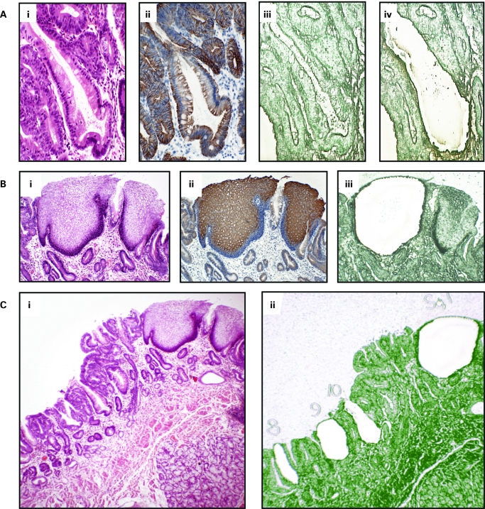
Figure 1. Laser capture microdissection. A(i) Individual crypts were identified using haematoxylin & eosin (H&E) slides A(ii) Cytokeratin 7 immunostaining of serial sections showing glandular differentiation of columnar lined oesophagus. A(iii), (iv) Individual crypts were microdissected from serial sections stained with methylene green and mounted on laser capture slides. B(i) Squamous islands were identified histologically using H&E slides. B(ii) Cytokeratin 13 immunostaining of serial sections showing mature squamous cell differentiation. B(iii) Squamous island after laser capture showing selective dissection of only squamous tissue. C(i), (ii) Individual structures were dissected evenly from across the block and numbered.

Figure 1