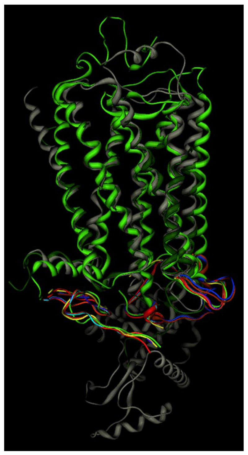
Figure 3.

Comparison of all available crystallographic GPCR structures. Rhodopsin structures 1F88 [53] (lt. green), 1HZX [76] (magenta), 1L9H [75] (yellow), 1GZM [103] (blue), 1U19 [52] (orange), 2HPY [74] (purple), 2G87 [74] (cyan), 2I35 [51] (rust) are superposed based on amino acid residues resolved in all structures, and shown only by the lt. green ribbon in these common areas to simplify the image. β2-adrenergic receptor structures 2RH1 [54] (grey) and 2R4R [57] (dk green) were superposed on the rhodopsin structures based on residues in the first three transmembrane domains.
