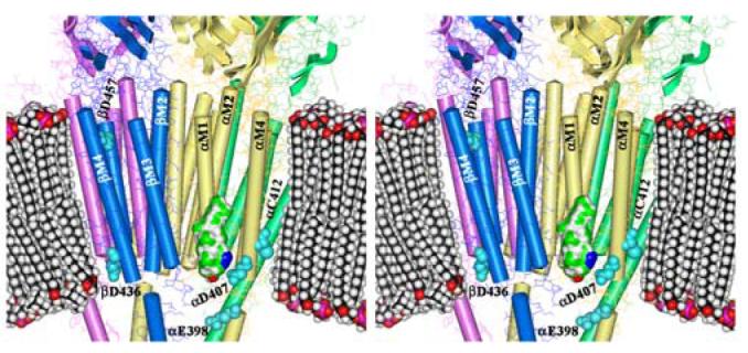
Figure 10. AziCholesterol Sites of Photoincorporation in the Torpedo nAChR Structure.

Shown is a stereo figure of the Torpedo nAChR cryo-electron microscopy structure (15; pdb #2BG9) focusing on the membrane spanning region. The α subunits are yellow, β is blue, γ is green, and δ is magenta. The pdb-designated α-helices and β-sheets are shown. A space-filling model of a phospholipid bilayer is included for scale as is a Connelly surface model of AziCholesterol with the Azide colored blue. Residues identified as labeled by [3H]AziCholesterol in the α, and β subunits are shown as cyan spaced-filled amino acids.
