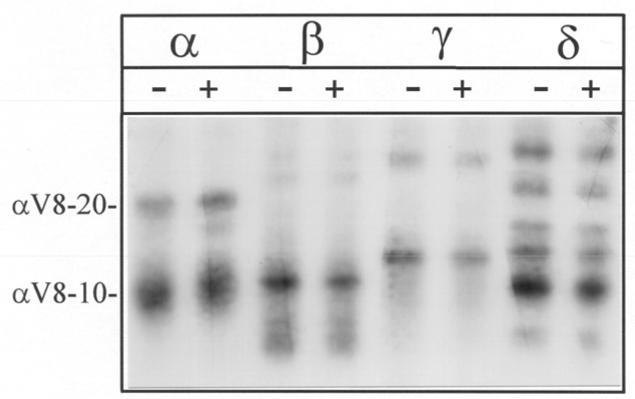
Figure 3. Proteolytic mapping of the sites of [3H]Azicholesterol incorporation into nAChR subunits using “in-the-gel” digestion with S. aureus V8 protease.
nAChR-enriched membranes were labeled with 1.25 μM [3H]Azicholesterol in the absence and in the presence of 400 μM carbamylcholine. After photolysis (365 nm for 10 min), membranes were resolved by SDS-PAGE (1.0 mm thick, 8% acrylamide). nAChR subunit bands were excised following identification by staining (Coomassie Blue) and transferred to the wells of a 15% acrylamide mapping for digestion with S. aureus V8 protease. Following electrophoresis, the mapping gel was stained with Coomassie Blue and processed for fluorography (16-week exposure). The principal [3H]Azicholesterol labeled proteolytic fragments, following the nomenclature of (12), are: αV8-20 (Ser-173- Glu-338); αV8-10 (Asn-339-Gly-437); βV8-22 (Ile-173/ Asn-183- Glu-383); βV8-12 (Met-384-Ala-469); γV8-24 (Ala-167/ Trp-170- Glu-372); γV8-14 (Leu-373/ Ile-413-Pro-489); δV8-20 (Ile-192- Glu-372); δV8-12 (Ile-192-Glu-280); δV8-11 (Lys-436-Ala-501).

