
Figure 4. Tryptic digestion of [3H]Azicholesterol labeled V8 protease fragments αV8-10, βV8-12, γV8-14, and δV8-11.
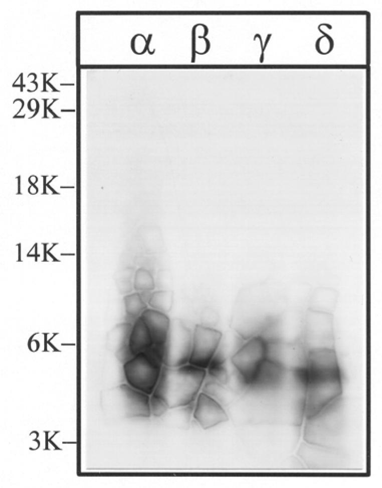
The V8 protease fragments αV8-10, βV8-12, γV8-14, and δV8-11, isolated from nAChRs labeled with 1.25 μM [3H]Azicholesterol in the absence of agonist, were exhaustively digested with trypsin (200% w/w) for four days. Aliquots of the total digests (∼5%) were fractionated by Tricine SDS-PAGE and then subjected to fluorography for 6 weeks. The migration of prestained molecular weight standards are indicated on the left. The principal band of 3H evident in each digest migrates with an apparent molecular mass of ∼ 5-kDa.