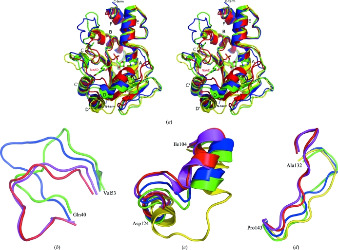
Figure 2.
(a) Overlay of apo BA NaMNAT (PDB code 3dv2, green; 2qtm, blue), apo BS NaMNAT (1kam, yellow) and BS NaMNAT–NaAD complex (1kaq, red; NaAD is shown in stick representation and colored by element). Secondary structures, N- and C-termini are labelled. (b–d) The three regions with the greatest conformational flexibility. Each region is enlarged and rotated to show most clearly the differences in conformation between the proteins. All structures shown in (a) plus BA NaMNAT–NaAD (PDB code 2qtr; magenta) are included. The first and last residues of each region are labelled for BA.

