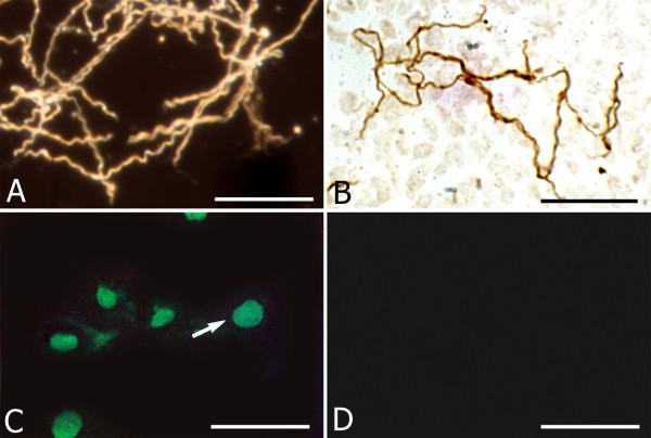
Figure 6.
Recovery of the typical vegetative form of spirochetes re-cultured in BSK II medium and nuclear fragmentation of rat primary astrocytes exposed to Borrelia burgdorferi. A: Dark field microscopy image of numerous Borrelia burgdorferi spirochetes (B31 strain) exhibiting the regular spiral form, re-covered in BSK-II medium following 1 week exposure to 5 mg Thioflavin S. B: Typical vegetative form re-covered from rat astrocyte culture exposed to Borrelia burgdorferi (ADB1) for 1 week, as revealed with a rabbit polyclonal anti-Borrelia burgdorferi antibody (BB-1017). Compare the regular spiral morphology of these spirochetes with those seen in Fig. 4H, where virtually all spirochetes showed atypical forms. C: Green fluorescent apoptotic nuclei of rat astrocytes as visualized with the TUNEL technique using FITC tagged dUTP. D: Uninfected primary astrocytes cultivated in parallel for 1 week did not show nuclear fragmentation. Bars: A, B: 25 μm; C, D: 50 μm.

