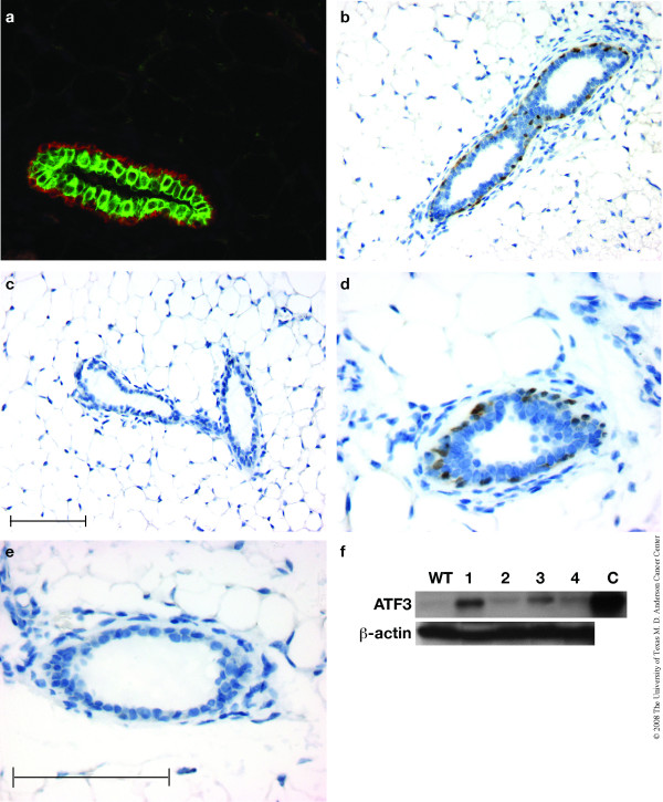
Figure 1.
ATF3 expression in myoepithelial cells of BK5.ATF3 mice. (a). A paraffin section (4 μm) of a mammary gland from an 8 week old BK5.ATF3 virgin female was stained with fluorescently labeled antibodies to CK5 (Texas Red, red) and CK8 (FITC, green) and examined by fluorescence microscopy. (b-e). Sections were also treated with a primary antibody to ATF3, then stained using horseradish peroxidase-coupled secondary antibody and a chromogenic substrate that produces a brown stain, and counterstained with hematoxylin. Mammary gland from a BK5.ATF3 transgenic female (b,d) was compared with a non-transgenic littermate (c,e). Scale bars = 100 μm; scale bar in panel c applies to panels b and c, scale bar in panel e applies to panels d and e. (f). Nuclear extracts were analyzed for ATF3 protein expression by immunoblotting as described in Materials and Methods. Equal amounts of protein from mammary glands of non-transgenic mice (lane WT) or BK5. ATF3 mice of line 1 (lane 1), line 2 (lane 2), line 3 (lane 3), or line 4 (lane 4) were analyzed. As a positive control, an extract from RAW 264.7 cells was analyzed (lane C). β-actin was used as loading control. An ATF3-specific band was detected in the 20–25 KD size range in the BK5.ATF3 mammary gland extracts and in the positive control extract; this band was barely detectable in extracts from non-transgenic mice (lane 1). The ATF3 band was abolished by preincubation with the ATF3 blocking peptide (not shown).

