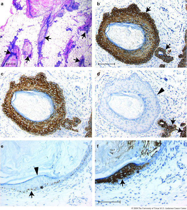
Figure 2.
Squamous metaplasia in virgin transgenic animals. Mammary glands from 21 nulliparous, BK5.ATF3 line 1 mice between 14 and 32 weeks of age and 18 age-matched, nulliparous non-transgenic littermates were examined histologically and by IHC. Transgenic mice from three other BK5.ATF3 lines that express the transgene were also examined (Table 1). (a) A low power view of multiple squamous metaplastic lesions in a single gland of a BK5.ATF3 line 4 female. (b-f) Two typical, cystic lesions from BK5.ATF3 line 1 mammary glands are shown, stained for (b) CK5; (c) CK6; (d) CK8; (e) ATF3; (f) CK10. Scale bar in a = 200 μm; scale bar in b = 100 μm, applies to panels b, c, and d; scale bar in f = 100 μm, applies to panels e and f.

