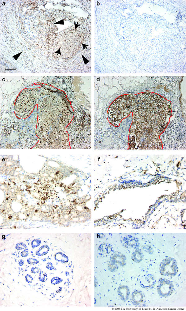
Figure 8.
Expression of ATF3 in human mammary tumors. Paraffin sections from human invasive ductal carcinomas that were less than 2 cm at presentation were examined by immunohistochemistry with an antibody directed against ATF3 (a-c,e,f). In (b), a blocking peptide for ATF3 (sc-188p, Santa Cruz) was added prior to the ATF3 antibody; the same region of the tumor is shown in (a) and (b). Panels a, c, and e represent tumors from three different patients. An adjacent section to that shown in (c) was analyzed for CK5 expression, and the corresponding region of the tumor is shown in (d). (f) shows a hyperplastic duct adjacent to an ATF3-positive tumor. (g-h), paraffin sections from normal mammoplasty tissues were examined by IHC with the same ATF3-specific antibody. Scale bar = 100 μm.

