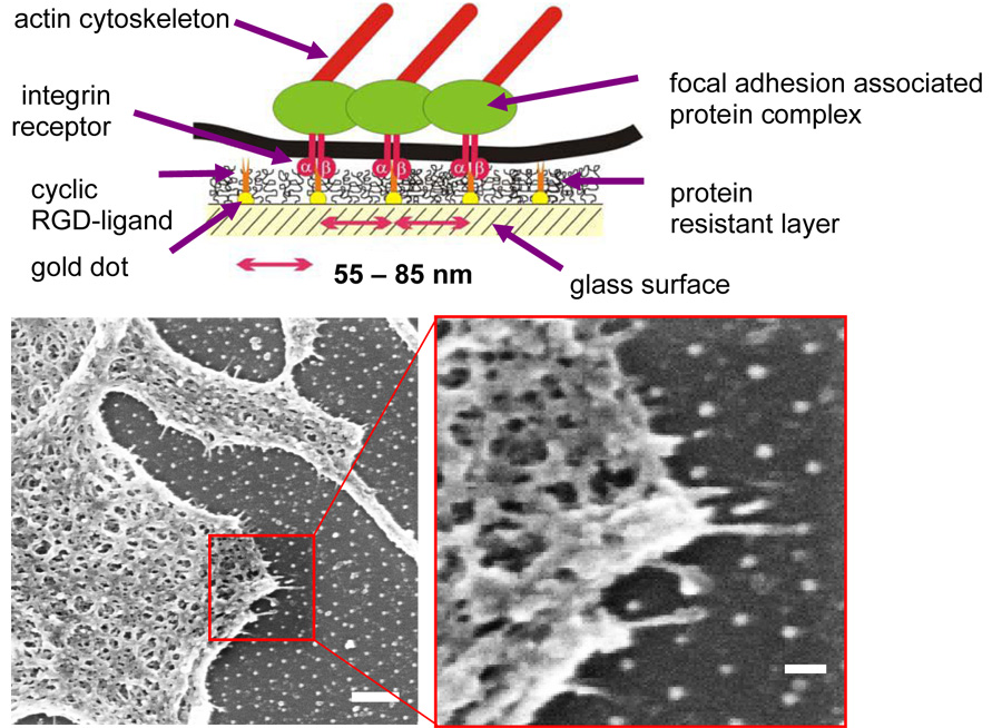
Fig. 2.
(Upper panel) Scheme of biofunctionalised nanopatterns to control integrin clustering (Arnold et al., 2004): gold dots are functionalised by c(-RGDfK-) thiols; glass areas between cell-adhesive gold dots are covalently bound to polyethyleneglycol to prevent unspecific protein binding. Therefore, cell adhesion is only mediated via c(-RGDfK-)-covered gold nanodots. (Bottom panels) Mc3t3 osteoblast in contact with a biofunctionalised 80-nm pattern and exhibiting cell protrusions sensing the pattern. Bars: 20 µm (left); 200 nm (right).

