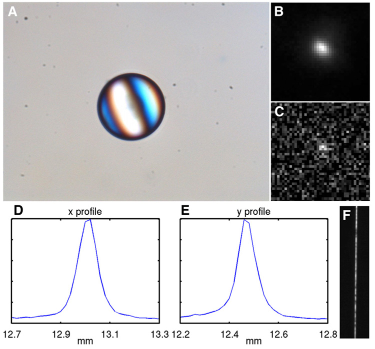
FIGURE 3.
(A) Micrograph of ion-exchange resin bead labeled with 99mTc-pertechnetate. Conversion-electron images of bead at 10-min exposure (B) and 2.5-s exposure (C). Profiles of conversion-electron image in x direction (D) and y direction (E). (F) Optical image of 10-µm slit using the same imaging components.

