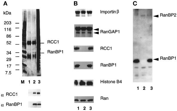
Figure 3.
Depletion of RanBP1 results in the codepletion of RCC1. (A) Anti-RanBP1 antibodies immunoprecipitate both RanBP1 and RCC1. Immunoprecipitates of anti-RanBP1 (lane 2) or preimmune sera (lane 1) were analyzed by SDS-PAGE together with 1 μl egg extract (lane 3). M indicates molecular weight marker. Top, a silver stained gel of the samples, with the position of RCC1 and RanBP1 indicated. Bottom, duplicate samples analyzed by Western blotting with anti-RCC1 and anti-RanBP1 antibodies. The faint band in lane 1 of the anti-RCC1 Western blot is the immunoglobulin heavy chain. (B) RCC1 is specifically and quantitatively codepleted with RanBP1. One μl of control (lane 1), immunodepleted (lane 2), and mock depleted (lane 3) cytosol were subjected to SDS-PAGE and Western blotting analysis with antibodies against importin β, RanGAP1, RCC1, RanBP1, histone B4, and Ran as indicated. (C) No RanBP1-like proteins remain in Xenopus egg cytosol after RanBP1 immunodepletion. One-microliter samples of control (lane 1), immunodepleted (lane 2), and mock-depleted extracts were subjected to SDS-PAGE and transferred to a PVDF membrane. The filter was incubated with [α-32P]GTP-bound GST-Ran to allow RanBP1 detection (see MATERIALS AND METHODS). RanBP1-depleted extracts show no low molecular weight Ran-binding proteins in this assay.

