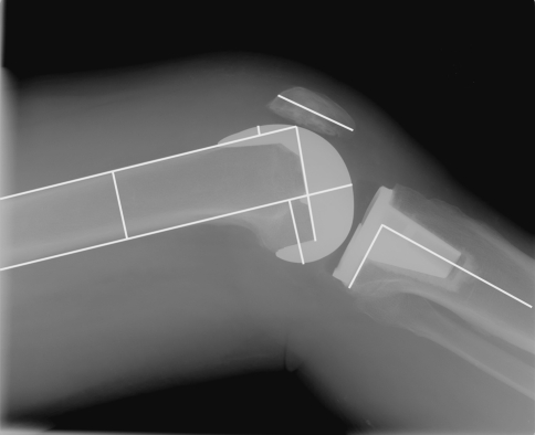
Fig. 2.
A true lateral postoperative radiograph demonstrates seven of the eight measurements recorded in each case: the same four measurements recorded preoperatively plus femoral component flexion relative to the anterior cortex, notching of the anterior cortex, and posterior slope of the tibial component. The eighth postoperative measurement was medial or lateral overhang of the femoral component at the distal joint line taken on the standing anteroposterior view.

