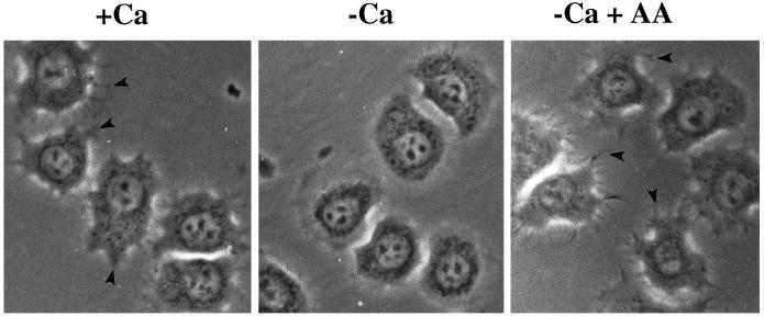
Figure 1.
Effects of extracellular Ca2+ on HeLa cell morphology during attachment and spreading on gelatin. HeLa cells were washed twice in PBS-Mg plus or minus Ca2+ before plating onto gelatin-coated polystyrene dishes at 37°C. One sample of cells resuspended in PBS-Mg minus Ca2+ was treated with AA at a final concentration of 1 μM before plating. After 30 min HeLa cells were observed by phase-contrast microscopy using a 40× phase-contrast objective. Arrowheads indicate filopodia seen when HeLa cells are spread in the presence of extracellular Ca2+ or in the absence of extracellular Ca2+ when treated with exogenous AA. Pictures are representative of five independent experiments.

