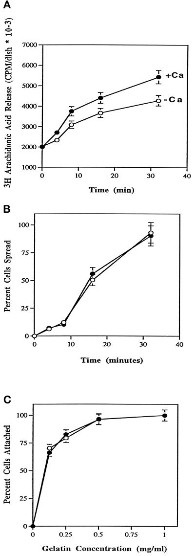
Figure 3.
Effects of extracellular calcium on release of AA (A), spreading rate (B), and attachment to various concentrations of gelatin (C). (A) HeLa cells at density of 2–3 × 10⁵ were labeled with [3H]AA at a final concentration of 0.25 μCi/ml for 12–16 h in the presence of serum. Cells were washed twice in PBS-Mg plus (•) or minus (○) Ca2+ and plated onto 60-mm gelatin-coated polystyrene dishes at a density of 2 × 10⁶ cells per dish. At the indicated times a 300-μl aliquot was taken from the supernatant and microfuged for 2 min. Three 40-μl fractions were then placed into scintillation vials for counting. Data are expressed as counts per minute per dish with SDs from four experiments. (B) At the indicated times, HeLa cells were assayed for percent cells spread from no less than three fields of view. Cells were spread in PBS-Mg plus (•) or minus (○) Ca2+. Data are represented with SDs from no less than five experiments. (C) HeLa cells were washed twice in PBS-Mg plus (•) or minus (○) Ca2+, treated with 1 mg/ml BSA, and plated at a density of 0.5 × 10⁵ cells per dish onto 35-mm polystyrene dishes coated with gelatin at the indicated concentrations. After 10 min at room temperature, cells were agitated for 5 s, the medium and unattached cells were decanted, and attached cells were scored. Data are expressed as percentage of cells attached and are normalized to cells attached at 1 mg/ml gelatin. Data are represented with SDs from at least five independent experiments.

