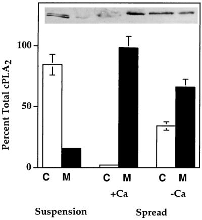
Figure 5.
Quantification of cPLA2 translocation from the cytosol to membrane during HeLa cell spreading on gelatin plus and minus extracellular Ca2+. HeLa cells were washed twice in PBS-Mg plus Ca2+ and plated onto 100-mm gelatinized dishes at a density of 8 × 10⁶ cells per dish, or cells remained in suspension. After 15 min the excess medium was removed, and cells were washed twice with PBS-Mg containing 2 mM EGTA. Cell lysates were separated into a soluble cytosolic fraction (C) and an NP-40 solubilized membrane fraction (M). Sixty micrograms of protein from each sample were electrophoresed per lane, and the blots were probed with mouse anti-human cPLA2 IgG. The optical density of each band was used to calculate the total amount of cPLA2 in each cytosol and membrane fraction. Data are expressed as a percentage of total cPLA2 for each treatment with SDs from five experiments.

