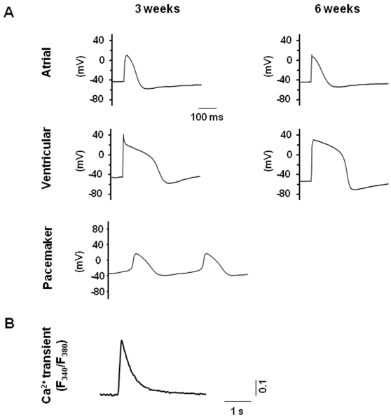
Figure 3. Electrophysiological recordings of hESC-CMs.
Action potential (AP) recordings from single cells were done using the whole-cell patch-clamp technique. hESC-CMs were categorized into pacemaker-, atrial- or ventricular-like phenotypes, based on such common electrophysiological characteristics as the AP amplitude (mV), upstroke velocity (mV/ms), APD50 and APD90 (ms), as well as the resting membrane potential (RMP, mV). (a) Representative ventricular-, atrial- and pacemaker-like action potentials, demonstrating electrophysiological heterogeneity in our hESC-CM population, and (b) Ca2+ transients recorded from hESC-derived cardiomyocytes, confirming calcium influx of these cells. See Materials and Methods for description of experimental parameters.

