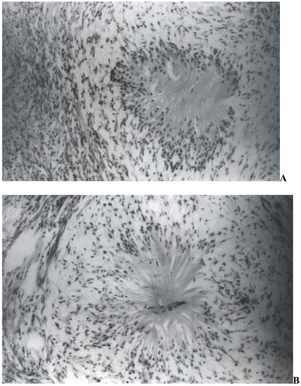
Figure 2.
(a): Section shows presence of rosette like structures between areas of typical neurofibroma. Rosettes display central eosinophilic, fibrillary core and peripheral palisading by neuronal cells. (H&E ×100) (b): Another rosette showing presence of central capillary in the fibrillary core. (H&E ×400).

