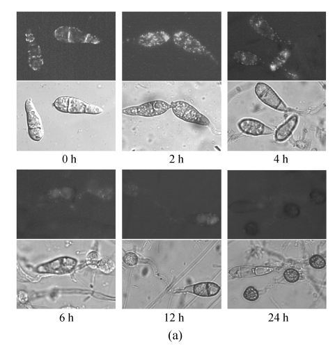
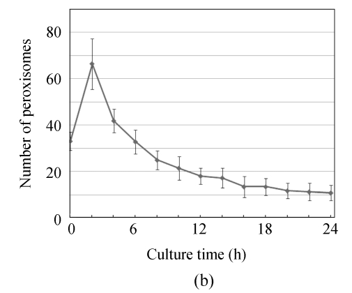
Fig. 5.
Numerical dynamic of peroxisome during appressorium differentiation. Conidia from GFPA transformants were allowed to form appressoria on hydrophobic membrane; (a) Fluorescence of different time points (the top, fluorescent image; the bottom, bright image); (b) Numbers of peroxisomes (fluorescent puncta) of different time points


