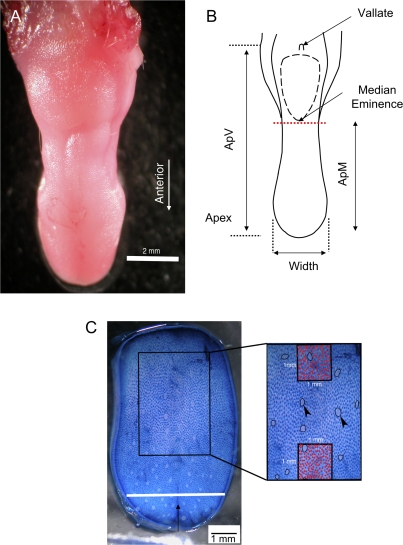
Figure 1.
Mouse tongue morphology. (A) Photomicrograph of a mouse tongue. (B) Line drawing reconstruction of the tongue from (A) showing the location of the vallate papillae and median eminence. Measurements where ApV, ApM, and width were taken are indicated with solid-headed arrows. The dashed red line marks the location where the tongue was sectioned for weighing. (C) Photomicrograph of a methylene blue–stained tongue surface showing location of the region (rectangle) where fungiform and filiform papillae numbers were measured. Inset displays the 2 regions where filiform papillae (red dots) numbers were counted. Arrowheads show examples of fungiform papillae.

