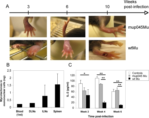
Figure 6. Mycolactone is present in the mononuclear cells of M. ulcerans-infected animals.
C57BL/6 mice (n = 3) were infected by footpad injection of 104 wtMu or mup045Mu bacilli. The distribution of mycolactone in PBMCs and in the mononuclear cell fraction of draining lymph nodes (DLNs), inguinal lymph nodes (ILNs) and spleens (Spleen) is shown 6 weeks post infection. Mononuclear cells were isolated from pooled samples of whole blood (1 ml), pooled DLNs (n = 3), or from the ILNs and spleens of 3 individual mice. Acetone-soluble lipid were then extracted from cell pellets and mycolactone concentrations determined by quantitative LC/MS-MS analysis. Means and SD are shown for ILNs and spleens. C) IL-2 production after whole blood stimulation with anti-CD3 and -CD28 antibodies for 24 h. Data are mean and SD of IL-2 concentrations, as measured in duplicate for pooled blood samples (n = 4) after 2, 4 and 6 weeks of infection, and are representative of three independent experiments. Differences in IL-2 concentration between groups were analyzed by one-way ANOVA (*: p<0,05; **: p<0,01).

