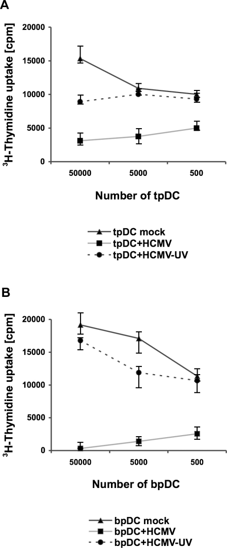
Figure 5. HCMV infection suppressed alloreactivity of pDCs in the MLR.
HCMV-infected (MOI 50) pDCs (pDC+HCMV) and pDCs incubated with UV-inactivated virus (pDC+HCMV-UV) were irradiated (30 Gray) at day five post infection. 5×10⁴–5×10² tpDCs (A) or bpDCs (B) were added as stimulator cells to 105 allogeneic purified CD4+ T cells (responder cells). As a control, mock-infected tpDCs or bpDCs (pDC mock) were included in each experiment. To measure T cell proliferation, cells were labelled with 1µCi 3H-thymidine per well at day five after coculture. The incorporation of 3H-thymidine (cpm) was measured for 18 h.

