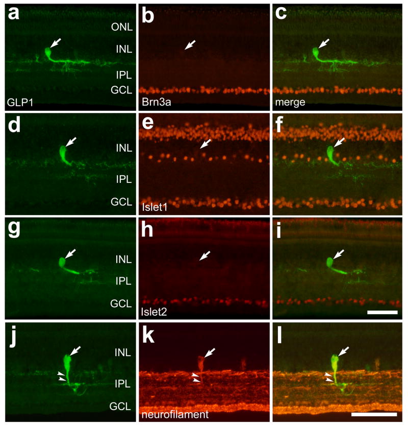
Figure 3.
Bullwhip cells express neurofilament, but do not express Islet1, Islet2 or Brn3a. Vertical sections of the ventral retina were labeled with antibodies to GLP1 (a, d, g and j) and Brn3a (b), Islet1 (e), Islet2 (h) or neurofilament (k). The micrographs in the column on the right are overlay images derived from the two panels to the left. The large arrows indicate GLP1-immunoreactive bullwhip cells and small arrows in panels j–l indicate the primary neurite of a bullwhip cell that is labeled for GLP1 and neurofilament. The calibration bar (50 μm) in panel i applies to panels a–c and g–i, and the bar in l applies to d–f and j–l. Abbreviations: ONL – outer nuclear layer, INL – inner nuclear layer, IPL – inner plexiform layer, GCL – ganglion cell layer.

