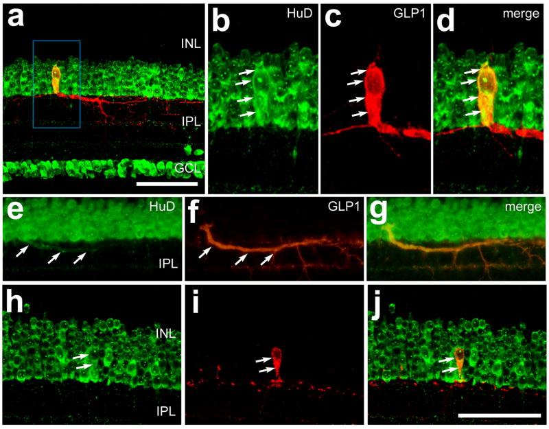
Figure 5.
Bullwhip cells and conventional glucagon-expressing amacrine cells (CGACs) express the RNA-binding protein HuD. Vertical sections of the retina were labeled with antibodies to HuD (green) and GLP1 (red). Arrows indicate a bullwhip cell labeled for GLP1 and HuD (b–d), HuD-immunoreactivity within the primary neurite of a bullwhip cell (e–g) or a CGAC that is immunoreactive for HuD and GLP1 (h–j). The images in panels a–d and h–j were obtained by using confocal microscopy and projection of four 1μm-thick optical sections. The images in panels e–g were obtained using epifluoresence microscopy. The calibration bar (50 μm) in panel a applies to panel a alone, and the bar in panel j applies to e–j. Abbreviations: INL – inner nuclear layer, IPL – inner plexiform layer, GCL – ganglion cell layer.

