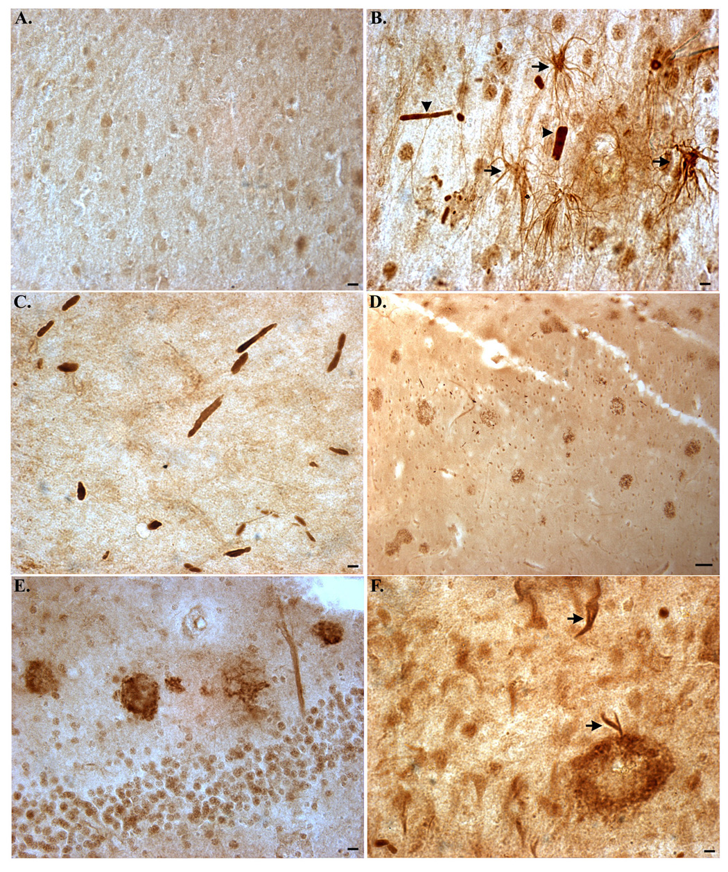
Figure 2. Detection of caspase-cleaved TDP-43 in the hippocampus of the Alzheimer’s disease brain.

(A): Representative age-matched control case with affinity-purified TDPccp (1:100) showing weak labeling within neurons of the hippocampus. (B–F): Representative staining with TDPccp in AD cases illustrating widespread labeling of the antibody within Hirano bodies (arrowheads, B and C), reactive astrocytes (arrows, B), within plaque-rich regions (low field, D; high field, E), and NFTs (arrows, F). All panels are representative staining in the hippocampus. Scale bars are equivalent to 10 µm except panel D, which represents 50 µm.
