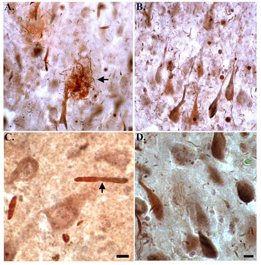
Figure 5. The TDPccp antibody co-localizes with an anti-ubiquitin mAB antibody in the AD brain.
Representative double immunostaining with TDPccp (red brown) and ubiquitin (blue gray) in the hippocampus of the AD brain. Note the co-localization of both markers within plaques (arrow, A), neurons with apparent NFT morphology (B and D), and Hirano bodies (arrow, C). Scale bars represent 10 µm.

