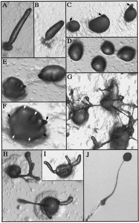
Figure 4.
Developmental morphology of ubcB-null cells plated on Na/K P04 agar plates. (A–B) Wild-type cells at the second finger/early culmination stage (18 h). (C) Wild-type tight aggregates forming tips (12 h), which are indicated by arrowheads. Note that each aggregate has only one tip. (D) ubcB-null cells at 18 h. Mounds first form at ∼8 h. Notice that no tips are present. (E) ubcB-null cells at 19 h as tips are first forming. (F) Enlargement of the lower left aggregate in E. The multiple tips are indicated by arrowheads. (G) ubcB-null cells plated at a higher density at 36 h. (H–I) ubcB-null cells at 36 h. (J) Mature wild-type fruiting body (26 h).

