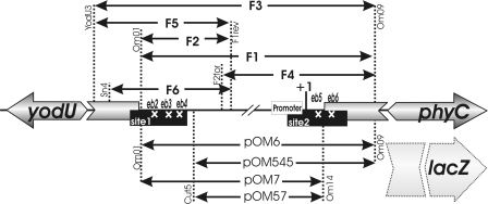
FIG. 1.
Schematic representation of the yodU-phyC intergenic region of B. amyloliquefaciens FZB45. The position of the phyCFZB45 promoter and the initiation point of phyC transcription (+1) are indicated. The two AbrB binding regions (sites 1 and 2) are indicated as filled boxes. The positions of the mutated binding areas (eb2, eb3, eb4, eb5, and eb6) within regions 1 and 2 are marked by white crosses. Double-headed arrows indicate DNA fragments amplified from parts of the entire phyC promoter region. DNA fragments used for lacZ fusions are listed at bottom, while DNA fragments used for DNase I footprinting, gel shift, and in vitro transcription are listed at top. DNA primers used for amplifying the respective DNA fragments are also shown at the vertical dotted lines.

