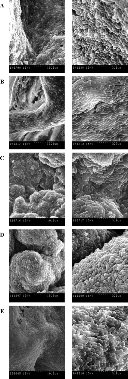
FIG. 3.
SEM images of rough colonies of M. chubuense (A), M. gilvum (B), M. obuense (C), M. parafortuitum (D), and M. vaccae (E). The right column shows the same sample as the left column but at greater magnification. These images are representative of the studies performed with six colonies of each species.

