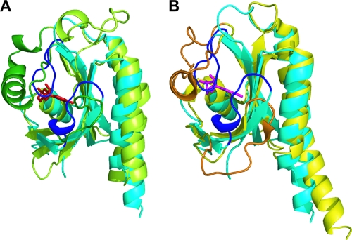
FIG. 6.
Structural comparison of the DevS GAF-B domain with GAF domains containing cyclic nucleotides. The structure of the GAF-B domain of DevS (cyan) is superimposed on the structures of the GAF-A domain (green) of adenylyl cyclase CyaB2 (A) and the GAF-B domain (yellow) of PDE2A (B). For the binding of cAMP (red) or cGMP (magenta), the α3-helix and the loop between the β2- and β3-strands of the GAF domains of PDE2A (orange) and CyaB2 (dark green) form the binding pocket. In GAF-B of DevS, two loops corresponding to the orange and dark green regions are presented in blue.

