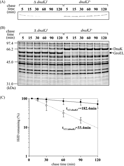
FIG. 5.
In vivo stabilities of HilD protein in wild-type cells and cells in which dnaKJ is disrupted. (A) The bacterial strains used were CS3659 (ΔdnaKJ) and CS3660 (dnaKJ+). Cells were grown to an OD600 of 0.5 at 30°C in L broth containing 500 μM IPTG to induce dnaKJ expression, followed by the induction of hilD expression by adding 0.005% arabinose for 30 min. Tetracycline (100 μg ml−1) and glucose (2%) were added, and samples were added to prechilled trichloroacetic acid (final concentration, 10%) at the indicated times. The proteins were separated on an SDS-10% polyacrylamide gel and then immunostained with anti-HilD antibody. (B) Coomassie brilliant blue-stained gel patterns of the same samples used for immunoblotting. (C) Quantification of the precipitated proteins relative to the value at 5 min. Mean values of at least three independent experiments are given. t1/2, half-life.

