Figure 3.
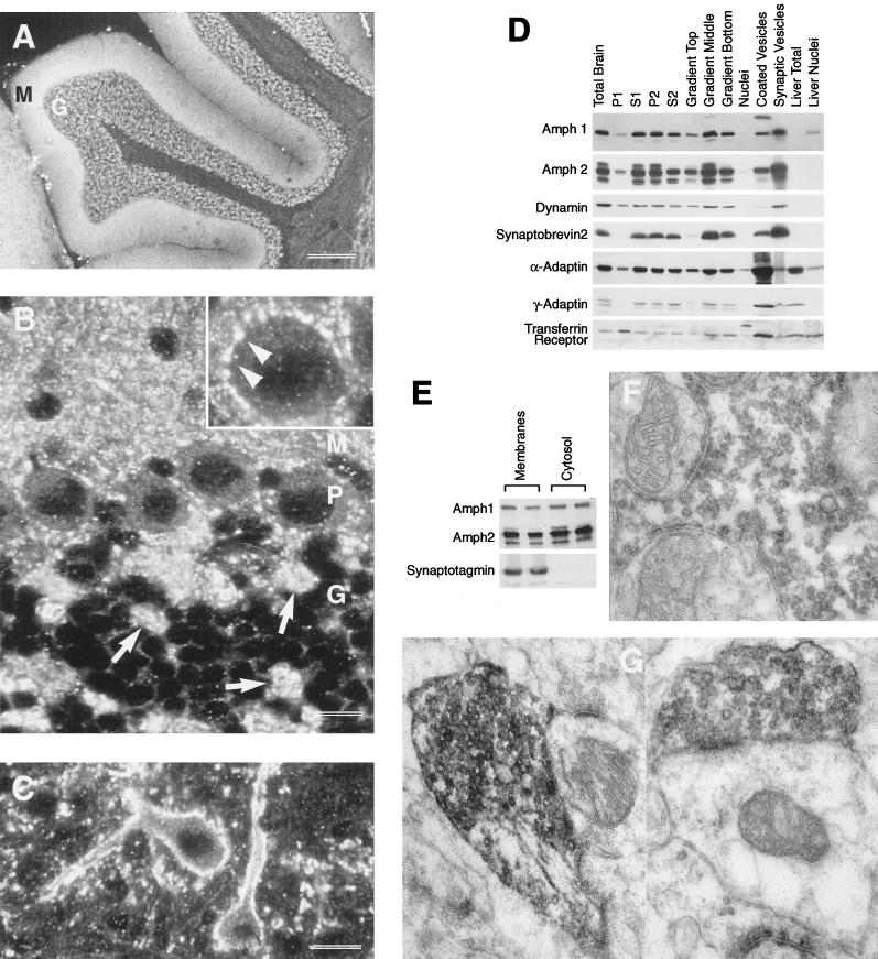
Subcellular distribution of Amph2 in rat brain. (A) By using Amph2 antiserum, the strongest staining for Amph2 is in the molecular (M) and granule cell (G) layers of the cerebellum. (B) At higher magnification, the presence of Amph in mossy fiber terminals of the granule cell layer (G) can be clearly seen (arrows). The Purkinje cell bodies (P) were surrounded by numerous small reactive terminals. This is more clearly seen in the inset (arrowheads). (C) Many cells in the pontine nucleus were densely innervated by fine Amph2 containing terminals. Bars: A, 300 μm; B, 14 μm; C, 9 μm. (D) Western blot analysis of subcellular fractions of rat brain. Subcellular fractionation was carried out according to MATERIALS AND METHODS. S1-P2 refer to successive pellets (P) or supernatants (S) of rat brain homogenized in 0.32 M buffered sucrose (see McMahon et al., 1992). The P2 fraction contains crude synaptosomes and was used for Percoll gradient separation into three predominant layers, the middle layer of which is enriched in isolated nerve terminals. Five micrograms of protein were loaded in each lane. (E) Amph1 and Amph2 both exist as membrane-associated and cytoplasmic pools. Synaptosomes were hypotonically lysed, followed by pelleting at 150 000 × g for 30 min to obtain a crude membrane fraction and cytosol, cleared of all synaptic vesicles (as shown by synaptotagmin control). Each fraction was loaded in duplicate, at 10 μg of protein per lane. (F and G) Electron micrographs showing the ultrastructural localization of Amph2 in the rat cerebellum. The large mossy fiber terminals in the granule cell layer were found to be heavily stained with Amph2 reaction product (F), as were climbing fiber terminals in the molecular/Purkinje cell layer (G). Within these structures, Amph2 staining was heavily concentrated on the outer layer of synaptic vesicles.

