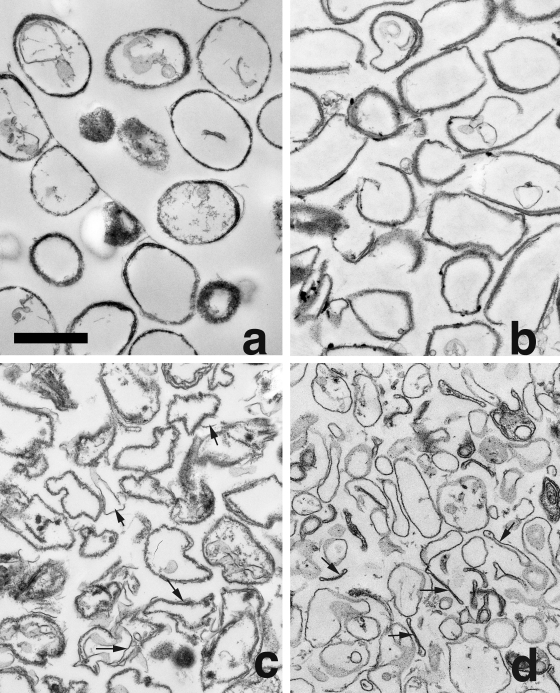
FIG. 4.
Electron micrographs of rind material generated by lysozyme and DNase digestion of spores with or without coat defects. Spores of strains PS533 (wild-type) (a), PS3328 (cotE) (b), PS4149 (gerE) (c), and PS4150 (cotE gerE) (d) were digested, and rind material was isolated, fixed, and examined by EM as described in Materials and Methods. Bars, 1 μm (all figures are at the same magnification). Arrows indicate thin rind material in gerE spores (c) and cotE gerE rinds that collapsed upon themselves (d).

