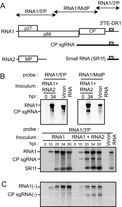
FIG. 1.
A small RNA is generated from the 3′-proximal region of RNA1 and packaged into virions in RCNMV-infected cells. (A) Schematic representation of viral RNAs of RCNMV. The genome is shown as horizontal lines with coding regions depicted as open boxes with the assigned viral protein designated. CP sgRNA and a small RNA (SR1f) are shown as thick horizontal lines below the genome diagram. The gray boxes indicate 3′TE-DR1. The regions covered by RNA probes are shown as thin arrows. MP, movement protein. (B) Accumulations of RNA1, CP sgRNA, and a small RNA (SR1f) in BY-2 protoplasts. Inoculated protoplasts were incubated at 17°C for the indicated times. Total RNAs extracted from protoplasts and purified virions were separated by gel electrophoresis and blotted onto a membrane. The top left, top right, and bottom panels show membranes hybridized with the DIG-labeled RNA probes specific for the 5′ UTR, middle region, and 3′ region, respectively. hpi, hours postinfection. (C) Accumulations of negative-strand RNA1 and CP sgRNA in BY-2 protoplasts. The same set of membranes shown in the bottom panel in panel B was hybridized with the DIG-labeled RNA probe specific for negative-strand RNA1.

