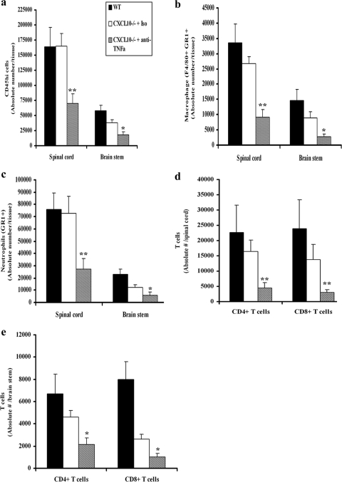
FIG. 3.
Macrophage, neutrophil, and T-cell infiltration into the CNS. Depo-Provera-treated WT and CXCL10−/− mice (n = 9/group) were infected with HSV-2 (2,000 PFU/vagina). On day 5 postinfection, 100 μg of anti-mouse TNF-α or isotypic control Ab (Iso) was administered intravenously (retro-orbitally) into HSV-2-infected CXCL10−/− mice. On day 7 postinfection, the mice were exsanguinated and the brain stem and spinal cord were removed from each mouse, processed, and analyzed for total infiltrating leukocytes (i.e., CD45HI) (a), activated macrophages (i.e., F4/80+ Gr1+) (b), neutrophil (c), CD3+ CD4+ T-cell or CD3+ CD8+ T-cell (spinal cord) (d), and CD3+ CD4+ T-cell or CD3+ CD8+ T-cell (brain stem) (e) content by flow cytometry (44). Bars represent the means ± SEM from three experiments. **, P values of <0.01; *, P values of <0.05 for comparison of the anti-TNF-α Ab-treated CXCL10−/− mice to the other two groups, as determined by ANOVA and Tukey's post hoc t test.

