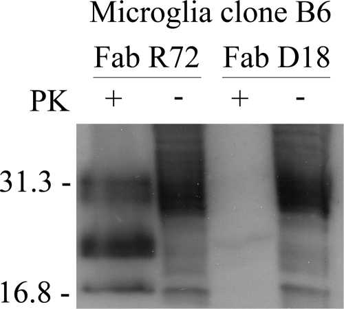
FIG. 4.
Immunoblot analysis of PrPSc-infected sheep microglial cell line after incubation with the recombinant anti-prion Fab D18. Primary sheep cultures were transformed with the SV40 virus large T antigen, cloned by limiting dilutions, and then inoculated with PrPSc. The clone that was positive for PrPSc was incubated with Fab D18 for 13 days and then cultured in the absence of the Fab for 4 weeks. Cells were lysed, treated with proteinase K (PK) (+) or not treated with proteinase K (−), and immunoblotted using the TeSeE Western blot kit (Bio-Rad, France). Lysates of replicate PrPSc-infected B6 cells that were treated with the control anti-prion Fab R72, which does not inhibit PrPSc accumulation, are included as a control for spontaneous loss of detectable PrPSc. The positions of molecular mass standards (in kilodaltons) are indicated to the left of the immunoblot.

