
FIG. 3.
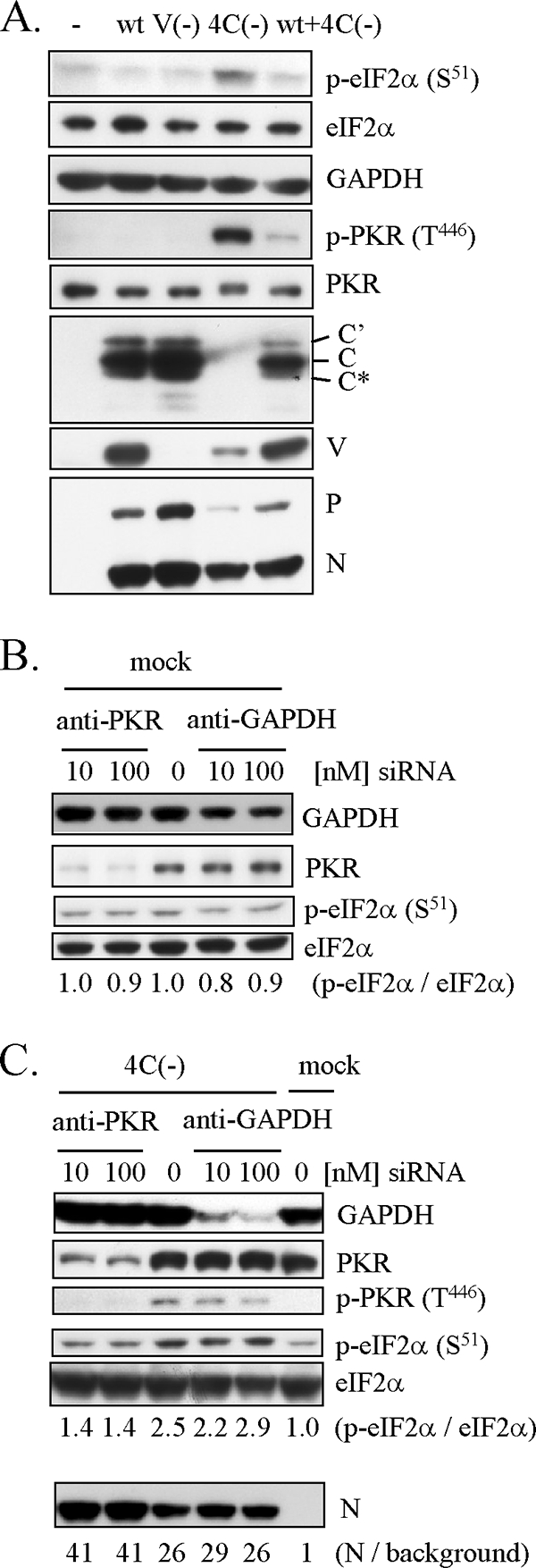
Phosphorylation of eIF2α and PKR in U118 cells infected with 4C(−). (A) U118 cells were mock infected (−) or infected with wt SeV, V(−), 4C(−), or a combination of wt SeV and 4C(−) and harvested at 30 h p.i. (B, C) U118 cells were transfected with increasing amounts of the siRNA specific for PKR or the control siRNA specific for GAPDH. At 40 h later, the cells were mock infected (B) or infected with 4C(−) (C) and harvested at 30 h p.i. (A to C) Proteins (10 μg) were analyzed by Western blotting with anti-V, anti-C, anti-SeV serum, anti-p-eIF2α (S51), anti-eIF2α, anti-p-PKR (T446), anti-PKR, or anti-GAPDH antibody. Ratios of p-eIF2α to eIF2α (B, C) and ratios of N to background (C) are also shown.