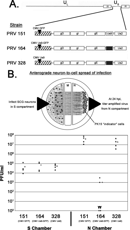
FIG. 2.
Dropping the EGFP tag from Us9 restores anterograde, neuron-to-cell spread in vitro. (A) Schematic of the PRV strains 151, 164, and 328. IR, inverted repeat. (B) SCG neurons were plated in the S compartment and allowed to extend neurites into the N compartment for 2 weeks. Axons were guided by a series of grooves scratched into the surface of the tissue culture dish. After 2 weeks, a monolayer of indicator PK15 cells was plated on top of the axon termini in the N compartment. Cell bodies in the S compartment were then infected at an MOI of 10 with PRV 151, 164, or 328. Four chambers were used for each type of infection (closed symbols). At 24 hpi, medium and infected cells were harvested together from either the S or the N compartment. Total PFU/ml were determined for each chamber. The mean value for the four samples is denoted by the offset open symbol. Black arrowheads denote the two plates infected with PRV 164 that showed no anterograde, neuron-to-cell spread. M, methocellulose compartment.

