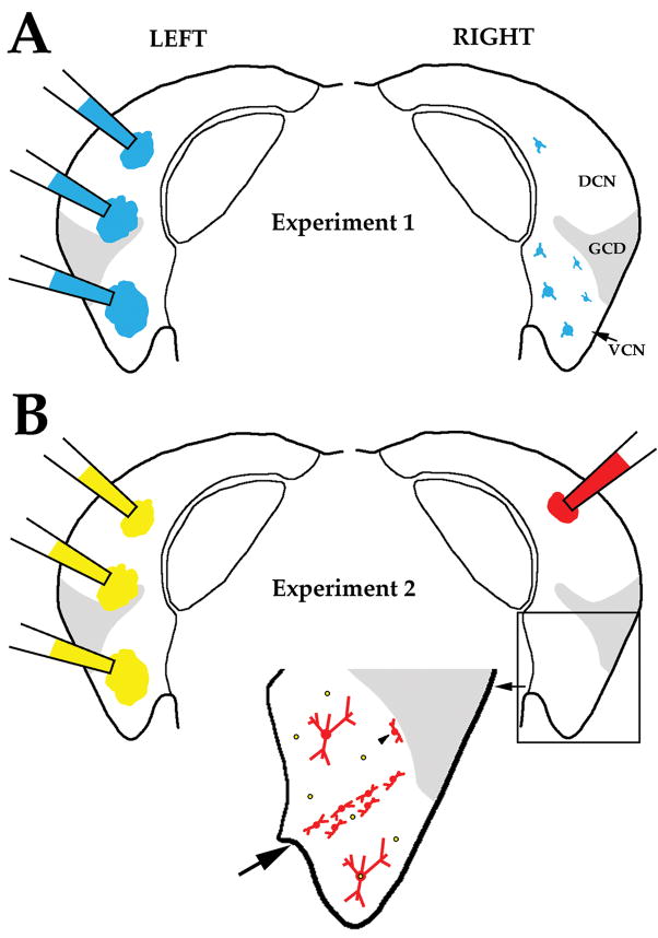
Figure 2.
Illustration of protocols and expected labeling patterns for the experiments performed in this study. Panel A and B each display coronal sections through the left and right CN. (A) In Experiment 1, FG or FB (depicted) was injected at several locations within the left CN to retrogradely label VCN multipolar cells in the right CN. Both tracers fill the soma (and proximal dendrites) of labeled VCN neurons, allowing us to characterize the size and location of their cell bodies. A few cells were labeled in the DCN but these were not analyzed. (B) In Experiment 2, DiY was injected at several locations within the left CN to retrogradely label VCN multipolar cells in the right VCN. DiY primarily labels the nucleus and labeled nuclei in the right VCN are depicted as yellow dots surrounded by black circles. In the same animal, a small injection of BDA was made in the right DCN. BDA is colored red because it was visualized with Cy3. BDA fills the soma and frequently the dendritic tree of labeled cells. Small BDA injections in the DCN produce a stripe of labeled planar multipolar cells in the corresponding frequency region of the ipsilateral VCN (large arrow). Labeled marginal multipolar cells (arrowhead) are observed adjacent to the granule cell domain. Two labeled radiate multipolar cells are shown –identified in this illustration by their large size and location outside the stripe of planar multipolar cells. Our hypothesis is that some radiate multipolar cells will also contain DiY (double labeled cell below stripe), whereas others will only contain BDA (single labeled cell above stripe). The number of double-labeled vs. single-labeled radiate multipolar cells provides information about whether all radiate multipolar cells project to both the ipsilateral DCN and the contralateral CN. By comparing the size distribution of double-labeled radiate multipolar cells in Experiment 2 with the size distribution of labeled multipolar cells in Experiment 1, we can test whether radiate multipolar cells are the sole source of the CN commissural pathway.

