Figure 3.
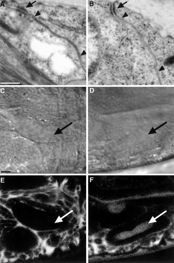
The ultrastructure and transepithelial barrier function of the septate junction is disrupted in the embryonic epithelia of coracle mutant embryos. (A and B) Transmission electron micrographs of the apical junctional complex in the epidermis of late stage 17 wild-type (A) and cor5 mutant (B) embryos. Although the adherens junction appears normal in the cor5 mutant tissue (black arrows; compare B with A), the region of the septate junction lacks the individual septae that characterize the pleated septate junction in the wild-type tissue (region between the arrowheads in A). (C–F) The transepithelial barrier function of the septate junction is disrupted in cor5 mutant embryos. (C–F) Differential interference contrast micrographs (C and D) and confocal optical sections (E and F) of stage 16 wild-type (C and E) and cor5 mutant (D and F) embryos showing the diffusion of a 10-kDa rhodamine–dextran injected into the hemocoel. Note that in the wild-type embryo the dextran fails to cross the salivary gland epithelium (arrows), whereas in the mutant embryo the dextran crosses the salivary gland epithelium as well as the epidermis. Bars, 250 nm (A and B), 10 μm (C–F).

