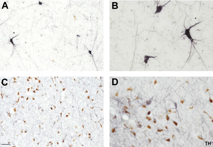
Figure 4. A–B, Immunohistochemistry for TH in the substantia nigra.

High power view (10× and 20× magnification respectively) of TH-ir neurons in the substantia nigra employing the citraconic anhydride solution. Note the enhancement in the intensity and morphology (somas more preserved and processes more delineated) compared to the conventional immunostaining method (C–D). Scale bar: A and C 100 µm; B and D 50 µm.
