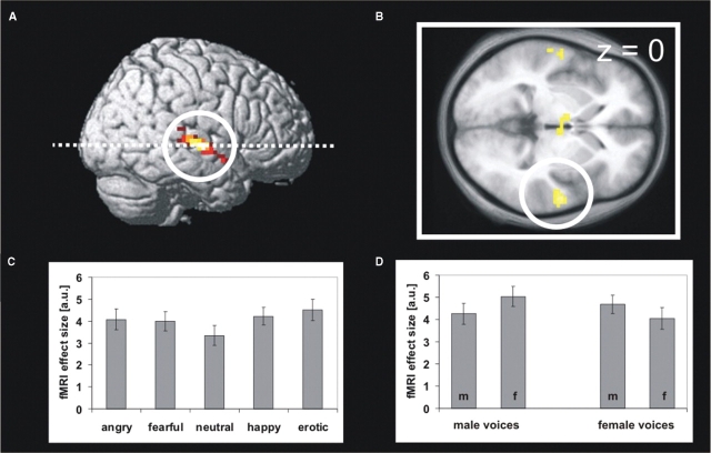
Fig. 2.
(A) Brain regions showing stronger activations to emotional than neutral prosody rendered on a right hemisphere of a standard brain and (B) a transversal slice (z = 0) of the mean T1 image obtained from the normalized brains of the fMRI participants. The activation cluster in the right mid STG is marked by a white circle. (C) Effect size of fMRI responses in right mid STG in arbitrary units (a.u.) for the five prosodic categories and (D) for erotic prosody dependent on gender of speaker and listener (m = male listener, f = female listener). Error bars represent standard errors of the mean.

