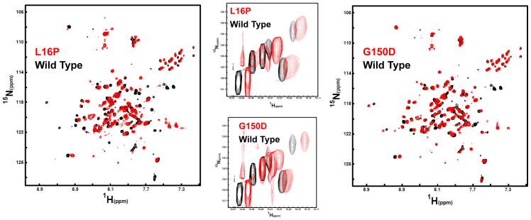
Figure 7.
Comparison of the NMR spectrum for WT PMP22 in TDPC micelles to the spectra of the Tr (G150D) and TrJ (L16P) mutants under identical conditions (25 mM acetate, pH 5.5, 150 mM NaCl, 0.2 % TDPC). All data were acquired at 800 MHz and 45°C. Left: The 1H,15N-TROSY spectrum of the L16P PMP22 mutant in red is overlaid on the spectrum of WT PMP22 protein in black. The L16P spectrum was collected using 128 increments and 248 scans per increment. The wild type spectrum was collected with 128 increments and 180 scans. Right: The 1H,15N-TROSY spectrum of the G150D PMP22 mutant in red is overlaid on the spectrum of WT PMP22 protein in black. Acquisition parameters for G150D were the same as those for L16P. Center: Mutant/WT TROSY spectral overlays for the indole ring 1H-15N from the protein’s 6 tryptophan residues (5 in PMP22 and 1 in the N-terminal tag).

