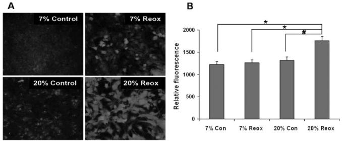
Fig. 4.
Nitrotyrosine immunostaining of cortical astrocytes 4 h after OGD. Astrocyte cultures were maintained at either 20 or 7% O2 both before and after OGD. Cells were stained using 3-NT antibodies after 4 h reoxygenation. A: Representative images of immunofluorescence. B: Quantitative comparison of immunocytochemical fluorescence. Relative fluorescence values were obtained as described in Materials and Methods and are expressed as the means ± SEM from n = 6–7 different experiments. Data were analyzed using one-way ANOVA with Holm-Sidak post hock test. *P < 0.01, #P < 0.05

