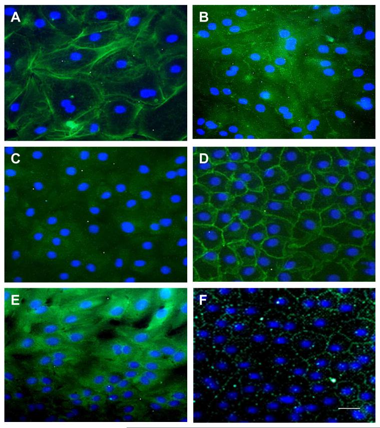
Figure 5. Maturaturation of Adherent Junction Components by In Vitro HCECs at Day 21.
Immunofluorescence staining of F-actin (by phalloidin) (A), VE-cadherin (B), E-cadherin (C), β-catenin (D), p190 (E), and ZO-1 (F) also showed gradual maturation. Negative control was similar to Fig. 4B (data not shown). The nuclear counterstaining is blue. The magnification bar represents 100 μm.

