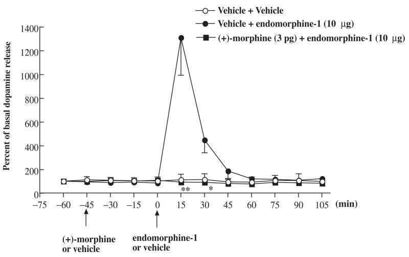
Fig. 6.
Effect of (+)-morphine on the extracellular dopamine release at the nucleus accumbens shell induced by endomorphin-1 microinjected into the ventral tegmental area in rats. Groups of rats were microinjected with (+)-morphine (3 pg) or vehicle into the ventral tegmental area 45 min before endomorphin-1 (10 μg) was microinjected into the ventral tegmental area. The perfusates from the microdialysis probe at the nucleus accumbens shell were collected every 15 min from 15 min before first microinjection and continuously collected for another 105 min after last microinjection. The perfusates were then analyzed their dopamine level. Each point represents the percent basal dopamine release and the vertical bar represents the S.E.M.; n = 3–9. Two-way ANOVA followed by Bonferroni post-test was used to test the difference between groups. For the groups of the rats injected with (+)-morphine and endomorphin-1 versus vehicle and endomorphin-1, Finteraction (7, 80) = 11.47, Ftreatment (1, 80) = 26.03, Ftime (7, 80) = 11.58, * P < 0.05, ** P < 0.005; only the data of the dopamine level after the 0 min time point were used for statistic analysis; the vehicle pretreatment followed by vehicle microinjected group was not included in the statistic analysis.

