Figure 3.
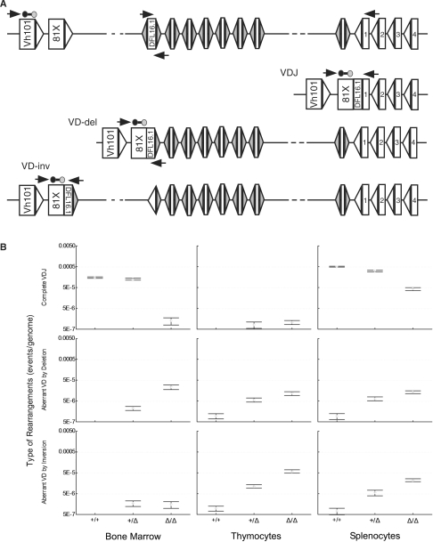
Frequency of complete VH-to-DH-to-JH and direct VH-to-DH rearrangements in cR2 and WT mice. (A) Diagram of a portion of the mouse IgH locus in its germline configuration (upper; only a subset of the total number of D segments bounded by RSS-12 are shown) as well as complete VDJ and direct VD rearrangements (VD-del and VD-inv, deletion and inversion) utilizing VH81x and DFL16.1. Real-time qPCR primers are shown as horizontal arrows and the probe (line bounded by circles). (B) DNA purified from the indicated tissues was analyzed for complete VDJ (upper) or direct VD joints (middle and lower; deletion or inversion). Hybrid joints (not illustrated) were excluded by placing the D segment primer just within that coding segment. Four animals from each of the three possible genotypes were examined and the mean values (bars) with SEs (whiskers) are shown.

