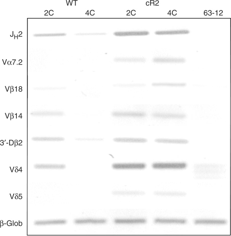
Figure 8.
Analysis of dsDNA RSS breaks in WT and cR2 thymocytes sorted based on DNA content (2C, 4C). Nuclei from several (pooled) 1-month-old mouse thymi were stained with PI and sorted into 2C (G0/G1) and 4C (G2/M) populations. Genomic DNA was subjected to LM-PCR to detect RSS breaks at the indicated antigen receptor gene segment RSSs. The negative image of an ethidium-stained agarose gel is shown. 63-12 is a RAG2-null cell line whose DNA served as a negative control. The direct amplification of β-globin serves as a DNA-loading control.

