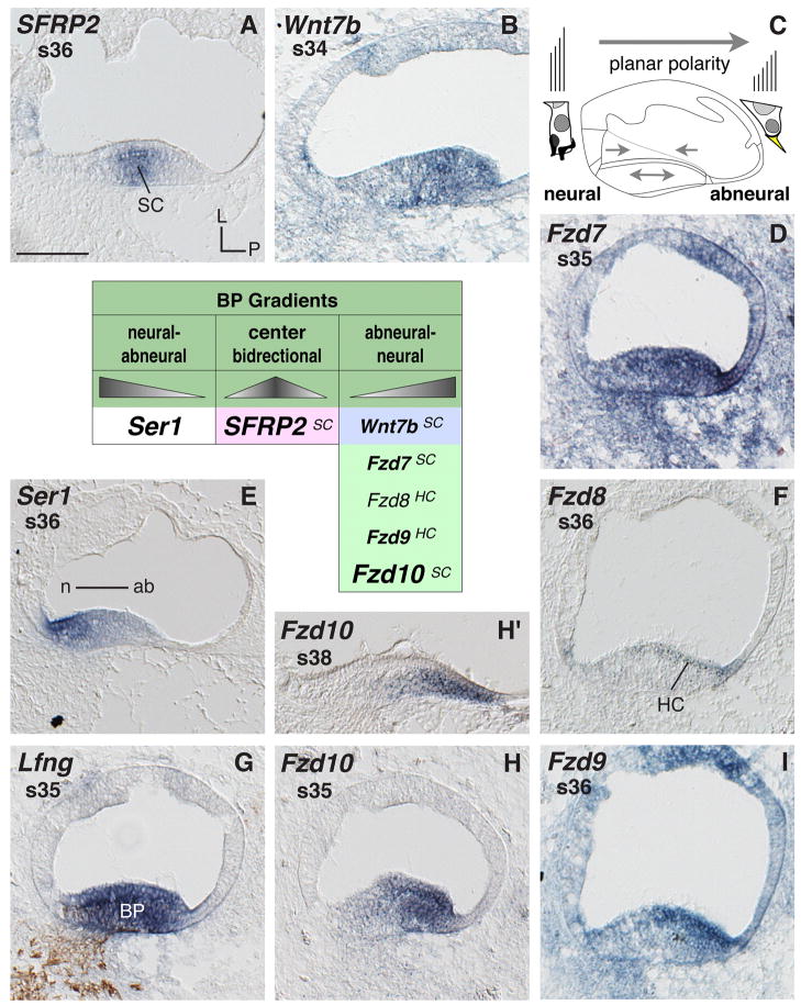
Fig. 4. Basilar papilla mRNA expression.
Gene expression in cross sections through the cochlear duct (CD) and basilar papilla (BP). A, B, D–F and H–I: BP expresses several genes locally concentrated across the neural-to-abneural axis. C: Cartoon cross section of the CD with enlarged neural vs. abneural BP hair cell schematics summarizes the main histological and physiological polarity across the BP (hair cell and bundle shape, innervation, and bundle orientation). Arrows within the CD indicate presumed directional signaling sources based on different gene expression. G: Double-labeling of the BP (Lfng probe) and of neurofilament (antibody 3A10). The table summarizes genes with locally concentrated expressions across the BP. Triangle shapes illustrate the relative signal levels of individual probes. A superscript at the listed gene refers to the main cell type expressing the transcript. Abbreviations: ab, abneural; BP, basilar papilla; HC, hair cells; L, lateral; n neural; P, posterior; s, stage; SC, supporting cells. Scale bar equals 100μm.

